
P071 T-cell, B-cell and monocytic infiltrations of myenteric plexus in IBD
Wiese, J.J.(1)*;Fascì, A.(1);Kühl, A.A.(1);Atreya, R.(2);Patankar, J.(2);Siegmund, B.(1);Prüß, M.S.(1);Schumann, M.(1);
(1)Charité - Universitätsmedizin Berlin, Department of Gastroenterology- Infectious Diseases and Rheumatology- Campus Benjamin Franklin, Berlin, Germany;(2)University Hospital Erlangen, Deptartment of Internal Medicine 1- Faculty of Medicine, Erlangen, Germany;
Background
Patients suffering from IBD frequently suffer from chronic visceral pain, severely affecting quality of life. To better understand factors impacting pain signaling by enteric nervous system (ENS) neuronal cells, we analyzed the infiltration of the myenteric plexus (MP) of the ENS by inflammatory cell populations. In detail, we characterized (i) the immune cell populations infiltrating the MP, (ii) neurotransmitter expression by MP cells and (iii) neuronal cell survival of MP in Crohn’s disease (CD) and ulcerative colitis (UC).
Methods
We included 12 UC and nine CD patients that received colectomies or ileocecal resections, respectively, and 10 control patients with surgeries for non-inflammatory reasons. FFPE material was used for immunohistochemistry analysis of the MP. Immunostaining was performed for the detection of ENS neurons (beta-III tubulin, PGP9.5), for T-cells (CD3, CD4, CD8 and FOXP3), for B-cells (CD20), for monocytes (CD68, CD163) and apoptosis (Annexin V) and the neurotransmitters CGRP and substance P. Intraganglionic and periganglionic immune cell populations were determined by conventional LSM confocal microscopy.
Results
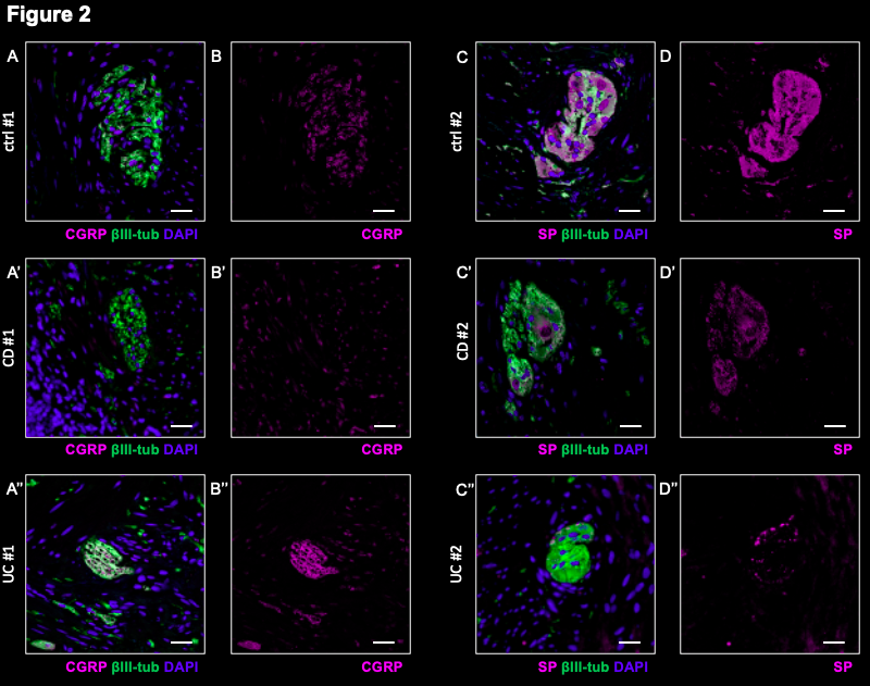
CD3+ and CD8+ intraganglionic T-cells for CD (CD3+: 1.7 ± 0.6 cells/10.000 µm²; CD8+: 4.8 ± 1.1 cells/10.000 µm²) were found to be increased compared to control patients (CD3+: 0.2 ± 0.1 cells/10.000 µm², p=0.04; CD8+: 0.4 ± 0.2 cells/10.000 µm², p=0.006), Figure 1. CD20+ B-cells and CD68+ monocytes were significantly increased in CD MP as well (CD20+ B-cells in CD: 0.8 cells/10.000 µm², CD20+ B-cells in controls: 0 cells/10.000 µm, p=0.02), CD68+ monocytes in CD: 12.3 cells/10.000 µm², CD68+ monocytes in controls: 3.5 cells/10.000 µm², p=0.001). CD8+ cytotoxic T-cells infiltrating the periganglionic area of the MP were significantly increased for UC. 1.6-fold increased expression of the neurotransmitter CGRP were detected for UC MP, p=0.01. Substance P was found to be 0.7-fold reduced for CD MP, p=0.01. MP-cell survival (Annexin V) was found to be reduced for UC MP, p=0.048. Figure 2 shows representative neurotransmitter immunostainings in two patients per condition.
Conclusion
In conclusion, MP immune cell infiltrations in CD are comprised of CD3+, CD8+ T-cells as well as CD68+ monocytes and CD20+ B-cells. Specifically periganglionically, CD8+ T-cells are found in UC. The expression of the neurotransmitter CGRP is increased in CD-MPs whereas levels of substance P are reduced in UC MP. UC-affected MPs revealed higher levels of apoptosis.