
P095 Anti-inflammatory effect of high acetate concentration on organoid-derived epithelial monolayer from patients with Ulcerative Colitis
Deleu, S.(1);Arnauts, K.(1,2);Machiels, K.(1);Huys, G.R.B.(3,4);Thevelein, J.(5,6);Raes, J.(3,4);Vermeire, S.(1,7);
(1)KU Leuven, Chrometa Targid, Leuven, Belgium;(2)KU Leuven, Development and Regeneration- Stem Cell Institute Leuven, Leuven, Belgium;(3)KU Leuven, Microbiology and Immunology- Rega Institute, Leuven, Belgium;(4)VIB, Center for Microbiology, Leuven, Belgium;(5)Erasmus High School, NovelYeast bv- Open Bio-Incubator, Brussels, Belgium;(6)KU Leuven, Molecular Cell Biology- Institute of Botany and Microbiology, Leuven, Belgium;(7)University Hospitals Leuven- KU Leuven, Gastroenterology and Hepatology, Leuven, Belgium; MIMOSA
Background
Short-chain fatty acids (SCFA), such as acetate and butyrate, have gained increasing interest for their potential to suppress IBD inflammation1,4. Most data have been generated for butyrate, showing beneficial effects on the gut microbiome composition, intestinal barrier function and immune system1. The effects of acetate are less known, despite its lower toxicity on epithelial cells and its ability to support growth of butyrate-producing bacteria by metabolic cross-feeding2. Furthermore, the probiotic effect of the biotherapeutic yeast Saccharomyces cerevisiae var. boulardii might be linked to its high acetic acid production3. Here, we studied the effect of acetate on inflammatory markers and barrier integrity using an organoid-derived epithelial monolayer system obtained from Ulcerative Colitis (UC) patients.
Methods
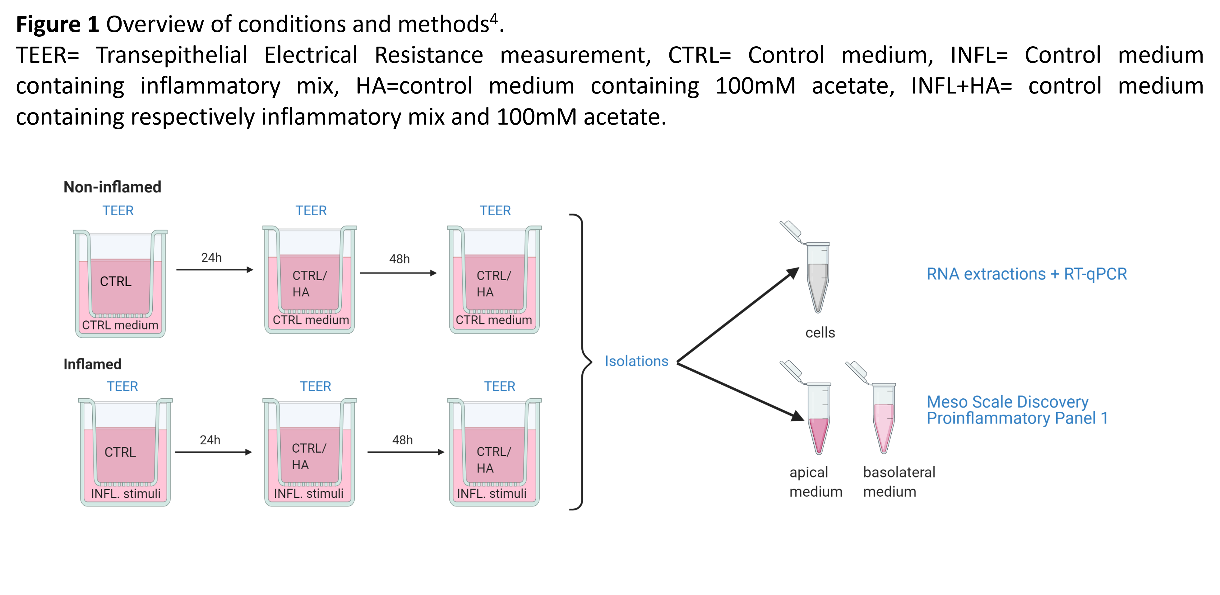
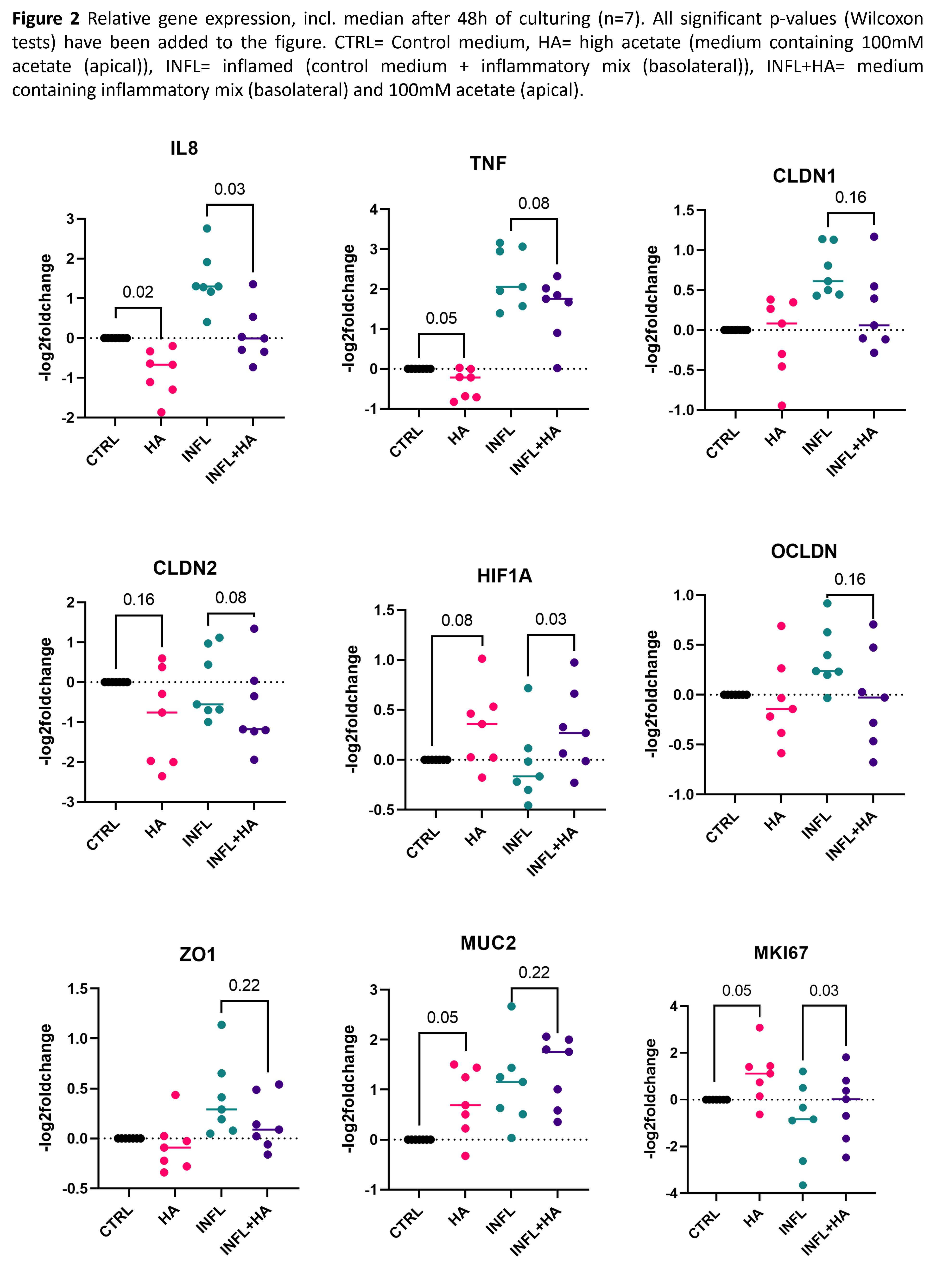
Confluent Transwell® organoid-derived monolayers evaluated by Transepithelial electrical resistance (TEER) of 8 UC patients were basolaterally stimulated with control medium (CTRL) or with an inflammatory mix (INFL) [100 ng/ml TNF-α, 20 ng/ml IL-1β, 1 µg/ml Flagellin] (Fig. 1). After 24h, the monolayer was stimulated apically with control medium or with high acetate (HA) [100mM] and TEER was measured at 0, 24, and 48h post-stimulation. After 48h, RNA extraction of the epithelial cell monolayers and qPCR for selected target genes (IL8, TNFα, CLDN1, CLDN2, HIF1A, ZO1, OCLDN, MUC2 and MKI67) was performed. Cytokine measurement was performed using Mesoscale Discovery (Proinflammatory panel 1) on apical and basolateral media. Data were analyzed using GraphPad Prism 9 (D'Agostino & Pearson test for normality followed by Wilcoxon tests).
Results
Transcriptomic analysis of the monolayer cells showed a decrease in expression of IL8 (p=0.03), TNFα (p=0.08) and increase of HIF1A (p=0.03) upon HA administration in inflamed conditions (Fig. 2) whereas the proliferation marker MKI-67 was upregulated (p=0,03). Moreover, these findings were supported by significant decreases in pro-inflammatory cytokines measured apically after HA stimulation (Fig. 3A) such as IFNγ, TNFα, IL6, IL8 and IL1β (all p=<0.01). Basolaterally, IL8 (p=0.02) and IL13 (p=<0.01) increased significantly upon HA stimulation (Fig.3B). Administration of HA was associated with a numerical though non-significant increase in TEER-values (p=0.15) (Fig. 4) suggesting a potential for improved barrier integrity.


Conclusion
In this UC patient-derived epithelial cell culture model, high acetate administration positively affected barrier integrity gene expression and TEER-values and suppressed expression and production of pro-inflammatory cytokines.
References:
1. Venegas, 2019
2. Gill, 2018
3. Offei, 2019
4. Deleu, 2021