
P198 Sonographic features of colonic pseudopolyposis in inflammatory bowel diseases
Silva Mendes, S.(1)*;Lepore, F.(2);Hussey, M.(3);Cataletti, G.(2);Maconi, G.(4);
(1)Braga Hospital, Department of Gastroenterology, Braga, Portugal;(2)Luigi Sacco University Hospital- University of Milan, Gastroenterology Unit, Milan, Italy;(3)University Hospital Galway, Department of Gastroenterology, Galway, Ireland;(4)Luigi Sacco University Hospital- University of Milan, Department of Gastroenterology, Milan, Italy;
Background
Colonic pseudopolyps are a frequent finding in inflammatory bowel disease (IBD). Yet there are no published data describing the characteristics of pseudopolyposis in intestinal ultrasound (IUS). This study aimed at identifying the key features of pseudopolyposis in IUS.
Methods
This case-control study included 8 patients with ulcerative colitis or Crohn’s colitis with extensive left colon pseudopolyposis and 24 matched IBD patients without pseudopolyps at colonoscopy. Luminal (diameters, thickening, stratification, margins, and vascularity) and intraluminal (vascular signals at color Doppler), and extraluminal (mesenteric fat) parameters of the left colon were compared. Anonymized still images and videos of these patients were blindly reviewed to estimate the accuracy in detecting this condition.
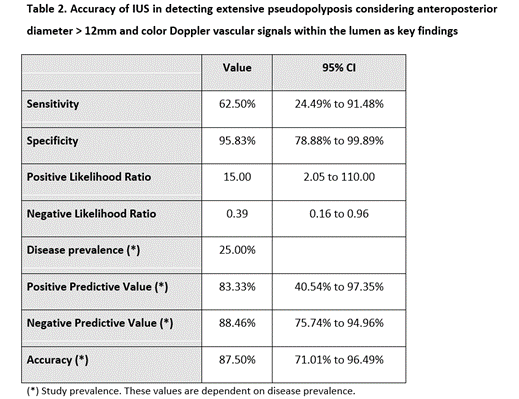
Results
Among the IUS parameters assessed, the anteroposterior diameter ≥ 12 mm and the presence of luminal vascular signals were significantly correlated with pseudopolyposis (table 1). The detection of both these findings were able to detect extensive pseudopolyposis with a sensitivity of 62.5% a specificity of 96.8%, a positive predictive value of 83.3% and negative predictive value of 88.46%, and an overall accuracy of 87.5% (table 2).

Conclusion
This is the first study describing the IUS features of pseudopolyposis in IBD. The potential use of IUS to assess pseudopolyposis might have an impact on IUS monitoring and surveillance of IBD patients with condition.